Anatomy of an Eye |
Anatomy, Physiology and |
Ted M. Montgomery, |
A short film, created in 1941, provides an excellent overview of the anatomy and physiology of the human eye. This black-and-white video is 11˝ minutes in length:
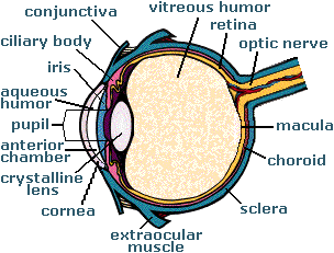
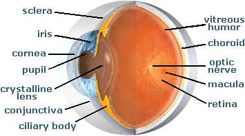
Click on a term in the eye graphics below to read a definition of it in the Glossary of Ocular Terms. From there, in some cases, you can link to more information.
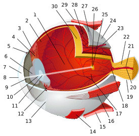
2 ora serrata 3 ciliary muscle 4 ciliary zonules 5 canal of Schlemm 6 pupil 7 anterior chamber 8 cornea 9 iris 10 lenticular cortex |
11 lenticular nucleus 12 ciliary process 13 conjunctiva 15 inferior rectus muscle 16 medial rectus muscle 18 optic disc 19 dura mater 20 central retinal artery |
21 central retinal vein 22 optic nerve 23 vorticose vein 24 bulbar sheath 25 macula lutea 26 fovea centralis 27 sclera 28 choroid 30 retina |
The eye manifests its refractive power via several curved surfaces, each separated by media with different indices of refraction. The most significant refractive surfaces are the anterior and posterior cornea and the anterior and posterior crystalline lens.
In the emmetropic eye (which has no refractive error), the range of corneal refracting power is between 39 and 48 diopters, while the range of lenticular refracting power is between 15 and 24 diopters. In the emmetropic eye, the axial length (from the posterior corneal surface to the retina) varies from 22 to 26 millimeters, or approximately an inch.
The following are the clear ocular media and structures (through which light passes before it reaches the retina) and their respective indices of refraction:
| • pre-corneal tear film • cornea • aqueous humor • crystalline lens • vitreous humor |
1.3375 1.3760 1.3360 1.4100 1.3360 |
Return to the Main Page ![]()
of Anatomy, Physiology & Pathology of the Human Eye
![]()
Copyright © 1998– by Ted M. Montgomery, O.D. Most rights reserved.